
Fichye:Cytokinesis-electron-micrograph.jpg
Cytokinesis-electron-micrograph.jpg (745 × 451 piksèl, gwosè fichye a : 200 kio, tip MIME li ye : image/jpeg)
Picture from English Wikipedia
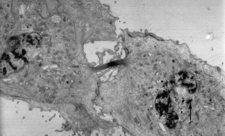
An electron micrograph image of a cell that has almost completed cell division and cytokinesis. Mitosis has already been completed. An arrow points to a centrosome still present near one of the nuclei.
From http://www.wadsworth.org/bms/SCBlinks/web_mit2/RES_MIT.htg/teleoph.jpg archive copy at the Wayback Machine, the Wadsworth Center, which is part of the New York State Department of Health and devoted to public education. Since it's part of the US government, I'll assume public domain.
Ce média est dans le domaine public des États-Unis d’Amérique car son auteur est l’administration américaine comme précisé dans le code fédéral au Titre 17, Chapitre 1, Section 105. Pour en savoir plus : droit d’auteur.
Attention : Ceci ne concerne que le travail du Gouvernement Fédéral et pas celui des États, ou d’une autre subdivision géographique ou politique du pays.
|
 | |
| Ce fichier a été identifié comme étant exempt de restrictions connues liées au droit d’auteur, y compris tous les droits connexes et voisins. | ||
Uploaded 07:21, 21 July 2005 .by user . Natalinasmpf . . 745x451 (87580 bytes) (An electron micrograph image of a cell that has almost completed cell division and cytokinesis. Mitosis has already been completed. An arrow points to a centrosome still present near one of the nuclei.
Ce média est dans le domaine public des États-Unis d’Amérique car son auteur est l’administration américaine comme précisé dans le code fédéral au Titre 17, Chapitre 1, Section 105. Pour en savoir plus : droit d’auteur.
Attention : Ceci ne concerne que le travail du Gouvernement Fédéral et pas celui des États, ou d’une autre subdivision géographique ou politique du pays.
|
| |
| Ce fichier a été identifié comme étant exempt de restrictions connues liées au droit d’auteur, y compris tous les droits connexes et voisins. | ||
)
Istorik fichye a
Klike sou yon dat/yon lè pou wè fichye a jan li te ye nan moman sa a.
| Dat ak lè | Minyati | Grandè yo | Itilizatè | Komantè | |
|---|---|---|---|---|---|
| Kounye a | 24 me 2011 à 12:54 | | 745 × 451 (200 kio) | Zephyris | Reverted to version as of 12:52, 24 May 2011 |
| 24 me 2011 à 12:54 |  | 745 × 451 (200 kio) | Zephyris | Reverted to version as of 12:50, 24 May 2011 Reversion seemed not to work | |
| 24 me 2011 à 12:52 |  | 745 × 451 (200 kio) | Zephyris | Reverted to version as of 12:50, 24 May 2011 Confusion with cached images | |
| 24 me 2011 à 12:52 |  | 745 × 451 (200 kio) | Zephyris | Oops, uploaded the original file last time by accident! | |
| 24 me 2011 à 12:50 |  | 745 × 451 (200 kio) | Zephyris | Inverted image: It is more common to show more intensly absorbing features in an electron micrograph (e.g. chromatin and the midbody) as dark rather than light. Asjusted levels and contrast: To both use the full histogram range and emphasise detail in the | |
| 1 desanm 2005 à 17:43 |  | 745 × 451 (86 kio) | Rasbak | Picture from English Wikipedia An electron micrograph image of a cell that has almost completed cell division and cytokinesis. Mitosis has already been completed. An arrow points to a centrosome still present near one of the nuclei. |
Itilizasyon fichye sa a
paj sa a itilize fichye sa a:
Itilizasyon global fichye a
Wiki sa a yo sèvi ak fichye sa a:
- Itilizasyon sou ar.wikipedia.org
- Itilizasyon sou bn.wikipedia.org
- Itilizasyon sou ca.wikipedia.org
- Itilizasyon sou en.wikipedia.org
- Itilizasyon sou es.wikipedia.org
- Itilizasyon sou gl.wikipedia.org
- Itilizasyon sou hy.wikipedia.org
- Itilizasyon sou it.wikipedia.org
- Itilizasyon sou kk.wikipedia.org
- Itilizasyon sou nl.wikipedia.org
- Itilizasyon sou nl.wikibooks.org
- Itilizasyon sou pl.wikipedia.org
- Itilizasyon sou pt.wikipedia.org
- Itilizasyon sou ru.wikipedia.org
- Itilizasyon sou sh.wikipedia.org
- Itilizasyon sou sl.wikipedia.org
- Itilizasyon sou sr.wikipedia.org
- Itilizasyon sou tr.wikipedia.org
- Itilizasyon sou uk.wikipedia.org